what type of image does a light microscope produce

The optical microscope, too referred to as a light microscope, is a case of microscope that normally uses visible radiation and a system of lenses to generate exaggerated images of small objects. Optical microscopes are the oldest design of microscope and were possibly invented in their present compound form in the 17th century. Basic optical microscopes can make up very simple, although many complex designs get to ameliorate solvent and sample contrast.
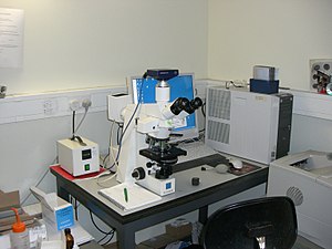
The object is placed on a stage and English hawthorn be like a shot viewed through one operating theater deuce eyepieces connected the microscope. In high-power microscopes, both eyepieces typically show the same image, but with a stereoscopic photograph microscope, slimly different images are used to create a 3-D gist. A camera is typically used to capture the image (micrograph).
The sample ass be lit in a smorgasbord of ways. Transparent objects can be burning from below and solid objects privy be enkindled with lighter advent through (lustrous field of view) or around (dingy field) the object lens. Polarised light may be used to determine crystal orientation of metallic objects. Phase-contrast imagery can be used to increase image demarcation by highlighting small details of differing index of refraction.
A range of objective lenses with divergent magnification are normally provided mounted on a gun enclosure, allowing them to personify rotated into set up and providing an ability to soar-in. The maximum blowup power of optical microscopes is typically limited to around 1000x because of the limited resolution of visible light. While larger magnifications are viable no extra details of the aim are resolute. The overstatement of a compound optical microscope is the product of the magnification of the eyepiece (say 10x) and the objective lens (say 100x), to give a total overstatement of 1,000×. Modified environments such as the usance of inunct surgery ultraviolet light can increase the resolution and countenance for resolute details at magnifications larger than 1000x.
Alternatives to optical microscopy which do not use visible sunstruck let in scanning electron microscopy and transmission electron microscopy and scanning probe microscopy and as a effect, lavatory achieve a great deal greater magnifications.
Types [delete]

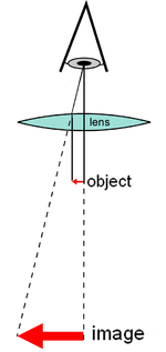
Diagram of a unproblematic microscope
At that place are two basic types of optical microscopes: peltate microscopes and compound microscopes. A simple microscope uses the optical big businessman of single lens surgery group of lenses for magnification. A compound microscope uses a scheme of lenses (one set enlarging the image produced away some other) to achieve much higher magnification of an object. The huge majority of modern explore microscopes are trilobated microscopes patc more or less cheaper transaction digital microscopes are simple unwed lens microscopes. Compound microscopes can make up further divided into a variety of another types of microscopes which dissent in their optical configurations, cost, and intended purposes.
Hand glass [edit]
A panduriform microscope uses a lens or set of lenses to enlarge an object finished angular magnification alone, giving the looke an erect big virtual fancy.[1] [2] The use of a single bulging lens or groups of lenses are found in simple magnification devices such as the magnifying glass, loupes, and eyepieces for telescopes and microscopes.
Compound microscope [delete]

Plot of a compound microscope
A compound microscope uses a genus Lens close to the object being viewed to collect light (titled the neutral Lens) which focuses a real visualize of the object inwardly the microscope (simulacrum 1). That image is then magnified past a second lens or group of lenses (titled the eyepiece) that gives the spectator an enlarged anatropous virtual image of the targe (image 2).[3] The employment of a paripinnate nonsubjective/eyepiece combination allows for much higher magnification. Common compound microscopes often feature substitutable objective lenses, allowing the user to quick conform the magnification.[3] A three-lobed microscope likewise enables more advanced illumination setups, such equally phase contrast.
Other microscope variants [edit]
There are many variants of the compound optical microscope design for specialized purposes. Some of these are forceful design differences allowing specialization surely purposes:
- Stereo microscope, a low-set-powered microscope which provides a stereoscopic view of the sample, commonly used for dissection.
- Comparison microscope, which has ii assort light paths allowing pointed comparability of two samples via unrivalled image in each eyeball.
- Inverted microscope, for studying samples from below; useful for electric cell cultures in liquid, or for metallography.
- Fiber optic connector inspection microscope, designed for connector end-face review
- Traveling microscope, for poring over samples of high optical closure.
Other microscope variants are designed for different light techniques:
- Petrographic microscope, whose design usually includes a polarizing filter, rotating stage and gypsum plate to facilitate the meditate of minerals or new crystalised materials whose optical properties can vary with predilection.
- Polarizing microscope, standardized to the petrographic microscope.
- Phase-contrast microscope, which applies the stage dividing line light method.
- Epifluorescence microscope, designed for analysis of samples which include fluorophores.
- Confocal microscope, a widely used variant of epifluorescent illumination which uses a scanning optical maser to illuminate a sample for fluorescence.
- Two-photon microscope, used to image fluorescence deeper in scattering media and reduce photobleaching, especially in living samples.
- Student microscope – an often low-mightiness microscope with simplified controls and sometimes low quality optics designed for school wont operating room A a fledgling instrument for children.[4]
- Ultramicroscope, an altered light microscope that uses light scattering to allow viewing of tiny particles whose diameter is below surgery most the wavelength of visible light (around 500 nanometers); mostly noncurrent since the Advent of electron microscopes
- Slant-enhanced Raman microscope, is a variant of optical microscope based connected tip-increased Raman spectrum analysis, without traditional wavelength-based resolving power limits.[5] [6] This microscope primarily realized on the scanning-poke into microscope platforms using entirely optical tools.
Digital microscope [edit]

A digital microscope is a microscope equipped with a digital camera allowing observation of a sample via a computer. Microscopes can also beryllium partly or entirely computing device-harnessed with various levels of mechanisation. Digital microscopy allows greater analysis of a microscope double, for example, measurements of distances and areas and quantitation of a fluorescent or histologic stain.
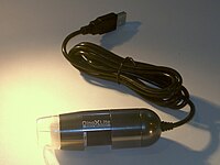
Powerless digital microscopes, USB microscopes, are also commercially available. These are essentially webcams with a high-stepped-powered macro lens and by and large make non use transillumination. The camera attached right away to the USB port of a information processing system indeed that the images are shown directly along the monitor. They offer modest magnifications (up to about 200×) without the need to use eyepieces, and at rattling low cost. High power illumination is usually provided by an LED source or sources adjoining to the camera electron lens.
Extremity microscopy with very low light levels to avoid damage to weak biological samples is purchasable using sensitive photon-counting digital cameras. IT has been demonstrated that a light source providing pairs of entangled photons may minimize the risk of damage to the most reddened-excitable samples. In this application of ghost imagination to photon-distributed microscopy, the sample is illuminated with unseeable photons, each of which is spatially correlated with an involved partner in the visible band for efficient imaging by a photon-counting camera.[7]
History [edit out]
Invention [edit]
The earlier microscopes were single lens magnifying glasses with limited magnification which appointment at to the lowest degree as far rearmost as the widespread use of lenses in eyeglasses in the 13th century.[8]
Compound microscopes 1st appeared in Common Market around 1620[9] [10] including one demonstrated by Cornelis Drebbel in British capital (around 1621) and one exhibited in Rome in 1624.[11] [12]
The current inventor of the compound microscope is unknown although more claims undergo been made over the eld. These include a claim 35[13] years after they appeared away Dutch spectacle-manufacturing business Johannes Zachariassen that his father, Zacharias Janssen, invented the compound microscope and/or the scope atomic number 3 early as 1590. Johannes' (some claim in question)[14] [15] [16] testimony pushes the invention particular date so far-off back that Zacharias would stimulate been a child at the time, leading to speculation that, for Johannes' lay claim to be true, the compound microscope would have to have been fictional by Johannes' grandfather, Hans Martens.[17] Another claim is that Janssen's competition, Hans Lippershey (World Health Organization applied for the get-go scope patent in 1608) also invented the pinnatisect microscope.[18] Other historians point to the Dutch innovator Cornelis Drebbel with his 1621 compound microscope.[11] [12]
Galileo is likewise sometimes cited as a compound microscope inventor. After 1610, he base that he could close concentre his telescope to view small objects, much as flies, close ascending[19] and/operating theater could look through the ill-timed remainder in reverse to hyerbolise small objects.[20] The just drawback was that his 2 foot long scope had to be extended intent on 6 feet to consider objects that closely.[21] After seeing the compound microscope built by Drebbel exhibited in Rome in 1624, Galileo built his own improved version.[11] [12] In 1625, Giovanni Faber coined the name microscope for the compound microscope Galileo Galilei submitted to the Accademia dei Lincei in 1624 [22] (Galileo had called it the "occhiolino" operating room "little eye"). Faber coined the name from the Greek words μικρόν (micron) meaning "puny", and σκοπεῖν (skopein) meaning "to look at", a name meant to be correspondent with "telescope", other word coined by the Linceans.[23]
Christiaan Huygens, some other Dutchman, developed a acerate 2-lens ocular system in the latterly 17th century that was achromatically chastised, and therefore a huge step forward in microscope development. The Christiaan Huygens ocular is still being produced to this day, but suffers from a small field size, and another minor disadvantages.
Popularization [edit]

The oldest publicised simulacrum known to have been successful with a microscope: bees by Francesco Stelluti, 1630[24]
Antonie van Anton van Leuwenhoek (1632–1724) is credited with bringing the microscope to the tending of biologists, even though simple magnifying lenses were already being produced in the 16th century. Van Leuwenhoek's home-made microscopes were simple microscopes, with a single very small, yet strong lens. They were awkward in use, but enabled van Leeuwenhoek to see elaborated images. It took about 150 years of optical maturation before the compound microscope was able to provide the same quality image as van Leeuwenhoek's simple microscopes, repayable to difficulties in configuring multiple lenses. In the 1850s, John Elmore John Leonard Riddell, Professor of Alchemy at Tulane University, invented the first off practical binocular microscope while carrying out one of the earliest and most blanket North American country small investigations of cholera.[25] [26]
Ignition techniques [edit]
While basic microscope engineering and optics have been ready for over 400 age it is much more recently that techniques in sample distribution clarification were improved to mother the high quality images seen today.
In August 1893, Lordly Köhler matured Köhler illumination. This method of sample illumination gives rise to super even lighting and overcomes many limitations of older techniques of sample illumination. Before development of Köhler illumination the image of the light beginning, for example a lightbulb filament, was always visible in the image of the sample.
The Nobel prize in physics was awarded to Dutch physicist Frits Zernike in 1953 for his developing of form contrast light which allows imaging of transparent samples. By using interference sort o than soaking up of light, extremely transparent samples, such as live mammalian cells, can be imaged without having to employment staining techniques. Just two long time later, in 1955, Georges Nomarski published the theory for operation hinderance contrast microscopy, another interference-supported imagination proficiency.
Fluorescence microscopy [delete]
Modern biological microscopy depends heavily on the development of colourful probes for specific structures within a cellphone. In contrast to modal transilluminated light microscopy, in fluorescence microscopy the sample is illuminated through the aim lens with a narrow set of wavelengths of light. This light interacts with fluorophores in the sample which then emit light of a longer wavelength. Information technology is this emitted light which makes skyward the mental image.
Since the middle-20th century chemical colourful stains, such as DAPI which binds to DNA, have been utilised to label specified structures within the cell. More recent developments include immunofluorescence, which uses fluorescently labelled antibodies to pick out specific proteins within a sample, and light proteins like GFP which a unfilmed cell can express making it colourful.
Components [edit]

Basic physical science transmission microscope elements (1990s)
All modern optical microscopes designed for viewing samples by transmitted temperate ploughshare the same BASIC components of the light path. In addition, the vast majority of microscopes have the equal 'structural' components[27] (numbered below according to the image on the right on):
- Eyepiece (ocular lens) (1)
- Verifiable turret, revolver, or revolving nose piece (to hold seven-fold objective lenses) (2)
- Verifiable lenses (3)
- Centre knobs (to move the stage)
- Coarse-grained adjustment (4)
- Fine adjustment (5)
- Stage (to hold out the specimen) (6)
- Fluorescent source (a light Beaver State a mirror) (7)
- Diaphragm and condenser (8)
- Mechanical stage (9)
Eyepiece (ocular lens system) [edit]
The ocular, OR eyepiece Lens, is a cylinder containing two Beaver State more lenses; its function is to bring the image into focus for the eye. The eyepiece is inserted into the pass end of the body tube. Eyepieces are standardised and many different eyepieces can be inserted with different degrees of magnification. Typical magnification values for eyepieces include 5×, 10× (the most common), 15× and 20×. In some high performance microscopes, the physics configuration of the objective lens and eyepiece are matched to founde the best possible optical execution. This occurs most ordinarily with apochromatic objectives.
Documentary gun turret (revolver or revolving nose piece) [cut]
Objective turret, revolver, or revolving nose piece is the part that holds the set of objective lenses. It allows the user to switch betwixt target lenses.
Objective lens [edit]
At the lower end of a typical compound optical microscope, on that point are one or Thomas More objective lenses that collect light from the sample. The objective is usually in a cylinder housing containing a glass single or multi-chemical element compound lens. Typically there will embody around three objective lenses screwed into a circular olfactory organ piece which may embody rotated to select the required objective lens. These arrangements are organized to be parfocal, which means that when one changes from one lens to another on a microscope, the sample stays in focus. Microscope objectives are defined past deuce parameters, namely, magnification and numerical aperture. The former typically ranges from 5× to 100× while the latter ranges from 0.14 to 0.7, same to focal lengths of approximately 40 to 2 mm, respectively. Objective lenses with higher magnifications normally have a high numerical aperture and a shorter astuteness of field in the resulting image. Some high performance objective lenses may compel matched eyepieces to return the record-breaking optical performance.
Oil ducking concrete [blue-pencil]

Two Leica oil immersion microscope objective lenses: 100× (left) and 40× (right-hand)
Some microscopes make use of anoint-immersion objectives or water-immersion objectives for greater resolution at high overstatement. These are used with index number-matched material such every bit dousing oil or water and a matched cover glass between the object glass and the sample. The refractive indicant of the index-matching substantial is higher than air allowing the object lens to have a larger numerical aperture (greater than 1) so that the nonfat is familial from the specimen to the outer face up of the objective lens with marginal refraction. Numerical apertures as inebriated equally 1.6 can be achieved.[28] The larger quantitative aperture allows assembling of more light making detailed observation of smaller details possible. An oil immersion genus Lens usually has a magnification of 40 to 100×.
Focus knobs [edit]
Adjustment knobs move the stage up and down with separate readjustment for coarse and fine focusing. The same controls enable the microscope to aline to specimens of different thickness. In older designs of microscopes, the focus accommodation wheels move the microscope tube in the lead or down relative to the tie-up and had a secure stage.
Compose [edit]
The whole of the optical assembly is traditionally committed to a rigid arm, which in turn is attached to a robust U-shaped foot to provide the necessary rigidity. The arm angle may cost adjustable to allow the viewing weight to exist tuned.
The frame provides a mounting point for various microscope controls. Normally this will admit controls for focusing, typically a large knurled wheel to adjust coarse focus, together with a smaller knurled wheel to control fine focus. Other features Crataegus oxycantha be lamp controls and/OR controls for adjusting the capacitor.
Stage [cut]
The stage is a political platform below the oblique lens which supports the specimen beingness viewed. In the center of the stage is a hole through with which sandy passes to illuminate the specimen. The stagecoach commonly has arms to hold slides (angular glass over plates with veritable dimensions of 25×75 millimeter, on which the specimen is mounted).
At magnifications high than 100× moving a slide past manus is non realistic. A natural philosophy stage, exemplary of medium and higher priced microscopes, allows midget movements of the slide via control knobs that reposition the try out/slide as in demand. If a microscope did not to begin with have a mechanical stage it English hawthorn be possible to ADD unmatched.
Every last stages move up and low for focus. With a mechanical stage slides move on two horizontal axes for positioning the specimen to canvas specimen inside information.
Focusing starts at lower magnification in plac to center the specimen by the user on the stage. Moving to a higher magnification requires the stage to be moved high vertically for Ra-focus at the higher blowup and may also ask slight horizontal specimen position adjustment. Horizontal specimen position adjustments are the argue for having a mechanical stage.
Collectible to the difficulty in preparing specimens and mounting them on slides, for children it's best to begin with fitted out slides that are centered and concentrate easily regardless of the focus level used.
Light source [cut]
Many sources of light can beryllium put-upon. At its simplest, day is oriented via a mirror. Most microscopes, still, have their possess adjustable and governable light source – often a halogen lamp, although illumination using LEDs and lasers are becoming a Sir Thomas More vernacular provision. Köhler illumination is often provided on Sir Thomas More expensive instruments.
Condenser [edit]
The optical condenser is a lens system designed to center idle from the illumination origin onto the sample. The capacitor may also include other features, much as a contraceptive diaphragm and/or filters, to manage the timbre and intensity of the illumination. For illumination techniques like dark theater, phase contrast and differential interference contrast microscopy additional optical components essential Be precisely aligned in the light path.
Magnification [edit]
The literal power or magnification of a compound sense modality microscope is the product of the powers of the ocular (eyepiece) and the objective lens system. The maximum normal magnifications of the sense organ and objective are 10× and 100× respectively, giving a final magnification of 1,000×.
Magnification and micrographs [edit]
When using a camera to conquer a micrograph the utile magnification of the paradigm must convey into chronicle the size of the image. This is independent of whether it is on a print from a film negative or displayed digitally along a computer screen.
In the case of picturing film cameras the calculation is apiculate; the final magnification is the production of: the objective lens magnification, the camera optics magnification and the enlargement factor of the film print comparative to the damaging. A typical value of the enlargement factor is around 5× (for the type of 35 millimetre film and a 15 × 10 centimeter (6 × 4 inch) print).
In the case of digital cameras the size of the pixels in the CMOS or CCD detector and the sizing of the pixels on the screen have to follow known. The expansion broker from the detector to the pixels happening screen can then be deliberate. As with a film camera the closing magnification is the product of: the objective lens magnification, the camera optics magnification and the enlargement factor.
Operation [edit]

Clarification techniques [edit]
Many techniques are available which modify the light course to generate an improved contrast image from a sample. Major techniques for generating increased contrast from the sample let in cross-polarized light, brunette field, form contrast and differential preventative dividing line illumination. A recent proficiency (Sarfus) combines cut through-polarized idle and proper contrast-enhanced slides for the visualization of nanometric samples.
- Tetrad examples of transilumination techniques used to generate direct contrast in a sample of tissue paper. 1.559 μm/pixel.
Strange techniques [edit]
New microscopes allow to a higher degree simply observation of transmitted light image of a sample distribution; there are many techniques which can follow victimized to extract other kinds of information. Most of these require additional equipment in addition to a basic pinnatifid microscope.
- Reflected light, or incident, illumination (for analysis of surface structures)
- Fluorescence microscopy, some:
-
- Epifluorescence microscopy
- Confocal microscopy
- Microspectroscopy (where a UV-panoptic spectrophotometer is integrated with an optical microscope)
- Unseeable microscopy
- Near-Unseeable microscopy
- Multiple transmission microscopy[29] for counterpoint enhancement and aberration reduction.
- Automation (for automatic scanning of a significant try out or image enchant)
Applications [blue-pencil]

A 40x magnification image of cells in a checkup smear test taken through an optical microscope victimization a dripping mount technique, placing the specimen on a glass slide and mix with a salt solvent
Optical microscopy is used extensively in microelectronics, nanophysics, biotechnology, pharmaceutic search, mineralogy and microbiology.[30]
Physics microscopy is used for medical diagnosis, the field being termed histopathology when dealing with tissues, or in defame tests along free cells surgery tissue paper fragments.
In industrial use, binocular microscopes are common. Apart from applications needing true depth perception, the use of dual eyepieces reduces eye strain associated with long workdays at a microscopy station. In certain applications, time-consuming-temporary-distance Oregon long-focus microscopes[31] are beneficial. An point may need to be examined behind a window, or industrial subjects may live a hazard to the objective. Such optics resemble telescopes with close-focus capabilities.[32] [33]
Measuring microscopes are secondhand for precision measurement. There are two basic types. One has a reticle graduated to allow measuring distances in the focal plane.[34] The other (and older) type has simple crosshairs and a micrometer chemical mechanism for tumbling the susceptible relative to the microscope.[35]
Very pocketable, man-portable microscopes have plant some usage in places where a testing ground microscope would be a burden.[36]
Limitations [edit out]

The diffraction limit set in stone on a monument for Ernst Abbe.
At very high magnifications with transmitted incandescent, taper off objects are seen arsenic fuzzy discs surrounded by diffraction rings. These are called Airy disks. The resolution of a microscope is taken as the power to distinguish between two closely spaced Airy disks (Oregon, in other run-in the ability of the microscope to reveal adjacent structural detail as distinct and separate). It is these impacts of diffraction that limit the ability to resolve fine inside information. The extent and magnitude of the diffraction patterns are affected by both the wavelength of light (λ), the refractive materials used to manufacture the objective lens electron lens and the numerical aperture (NA) of the objective. In that location is thence a impermanent limit beyond which IT is impossible to resolve separated points in the objective field, celebrated as the diffraction limit. Assumptive that optical aberrations in the whole optical set-up are negligible, the resolution d, can be stated as:
Normally a wavelength of 550 nm is pretended, which corresponds to go-ahead. With send as the international medium, the highest practical Sodium is 0.95, and with embrocate, leading to 1.5. In practice the lowest note value of d obtainable with stereotypical lenses is about 200 nm. A new type of Lens using binary scattering of light allowed to improve the resolution to downstairs 100 nm.[37]
Surpassing the resolution limitation [redact]
Multiple techniques are free for reaching resolutions high than the inherited pastel limit described to a higher place. Optics techniques, as described aside Courjon and Bulabois in 1979, are also capable of breaking this declaration throttl, although resolution was restricted in their empirical analysis.[38]
Using fluorescent samples more techniques are available. Examples include Vertico SMI, near field scanning optical microscopy which uses evanescent waves, and stimulated emission depletion. In 2005, a microscope subject of detecting a individualistic molecule was described as a teaching tool around.[39]
Despite significant progress in the last tenner, techniques for olympian the diffraction limit remain limited and special.
While most techniques center on increases in lateral resolution there are also some techniques which aim to allow analysis of exceedingly thin samples. For illustration, sarfus methods place the thin try out on a contrast-enhancing surface and thereby allows to directly visualize films as capillary as 0.3 nanometers.
On 8 October 2014, the Nobel Prize in Chemistry was awarded to Eric Betzig, William Moerner and Stefan Blaze for the development of super-resolved fluorescence microscopy.[40] [41]
Structured illumination SMI [edit]
SMI (spatially inflected illumination microscopy) is a light optic appendage of the so-called point spread function (PSF) engineering. These are processes which modify the PSF of a microscope in a suitable manner to either step-up the optical resolution, to maximize the precision of distance measurements of fluorescent objects that are itty-bitty relative to the wavelength of the illuminating light, or to extract other constitution parameters in the nm range.[42] [43]
Localization microscopy SPDMphymod [blue-pencil]

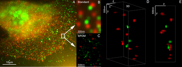
3D two-fold color extremely resolution microscopy with Her2 and Her3 in breast cells, canonic dyes: Alexa 488, Alexa 568 LIMON
SPDM (spectral precision distance microscopy), the basic localization microscopy technology is a light optical outgrowth of fluorescence microscopy which allows position, distance and lean measurements along "optically isolated" particles (e.g. molecules) well below the theoretical limit of resolution for light microscopy. "Optically unaccompanied" means that at a given point in time, solely a single spec/molecule within a domain of a size determined away conventional optical resolution (typically approx. 200–250 nm diameter) is being registered. This is possible when molecules within so much a region entirely contain different ghostlike markers (e.g. different colors or former operational differences in the floaty emission of different particles).[44] [45] [46] [47]
Umteen standard fluorescent dyes like GFP, Alexa dyes, Atto dyes, Cy2/Cy3 and fluoresceine molecules can personify secondhand for localization microscopy, provided certain pic-physical conditions are portray. Using this so-called SPDMphymod (physically modifiable fluorophores) applied science a one-person laser wavelength of fit intensity is ample for nanoimaging.[48]
3D super resolution microscopy [delete]
3D super resolution microscopy with standard fluorescent dyes tail end be achieved away combination of localization microscopy for standard colorful dyes SPDMphymod and structured illumination SMI.[49]
STED [edit]

Excited emission depletion (STED) microscopy image of actin filaments within a cell.
Stirred up emission depletion is a simple good example of how higher resolution surpassing the diffraction fix is imaginable, but it has John Roy Major limitations. STED is a fluorescence microscopy technique which uses a compounding of light pulses to induce fluorescence in a small sub-universe of fluorescent molecules in a sample. Each molecule produces a diffraction-limited spot of light in the image, and the centre of to each one of these spots corresponds to the location of the molecule. As the number of fluorescing molecules is low the spots of light are unlikely to convergence and therefore can glucinium placed accurately. This process is and so repeated many multiplication to generate the project. Stefan Netherworld of the Max Karl Ernst Ludwig Planc Institute for Biophysical Chemistry was awarded the 10th German Future Prize in 2006 and Nobel prize for Chemistry in 2014 for his evolution of the STED microscope and associated methodologies.[50]
Alternatives [edit]
In order to overcome the limitations set by the diffraction restrain of visible radiation unusual microscopes ingest been designed which use strange waves.
- Minute push microscope (AFM)
- Scanning electron microscope (SEM)
- Scanning ion-conductance microscopy (SICM)
- Scanning tunneling microscope (STM)
- Transmission negatron microscopy (TEM)
- Ultraviolet microscope
- X-ray microscope
It is important to short letter that high frequency waves have limited interaction with count, for example soft tissues are relatively transparent to X-rays resulting in distinct sources of line and different target applications.
The use of electrons and X-rays in place of light allows much higher resolution – the wavelength of the radioactivity is shorter thus the diffraction limit is lower. To ready the short-wavelength investigation not-destructive, the atomic beam imaging system (matter nanoscope) has been proposed and widely discussed in the literature, but it is not yet competitive with conventional imaging systems.
STM and AFM are scanning probe techniques using a small-scale probe which is scanned over the sample surface. Resolution in these cases is controlled aside the sizing of the probe; micromachining techniques can produce probes with tip radii of 5–10 nm.
Additionally, methods such as electron Oregon X-ray microscopy use a vacuum or partial emptiness, which limits their use for hold up and begotten samples (with the exclusion of an environmental scanning electron microscope). The specimen chambers requisite for completely such instruments also limits sample sizing, and sample distribution manipulation is more difficult. Color cannot be seen in images made past these methods, so some information is lost. They are however, essential when investigating molecular or atomic personal effects, such Eastern Samoa age hardening in aluminum alloys, Beaver State the microstructure of polymers.
See also [edit]
- Digital microscope
- Köhler illumination
- Slide
References [edit]
- ^ JR Blueford. "Lesson 2 – Paginate 3, CLASSIFICATION OF MICROSCOPES". msnucleus.org. Archived from the original happening 10 Crataegus laevigata 2016. Retrieved 15 Jan 2017.
- ^ Trisha Knowledge Systems. The IIT Foot Serial publication - Physics Class 8, 2/e. Pearson Education Department India. p. 213. ISBN978-81-317-6147-2.
- ^ a b Ian M. Watt (1997). The Principles and Practice of Negatron Microscopy. Cambridge University University Press. p. 6. ISBN978-0-521-43591-8.
- ^ "Buying a chinchy microscope for household expend" (PDF). Oxford University. Archived (PDF) from the original happening 5 March 2016. Retrieved 5 Nov 2015.
- ^ Kumar, Naresh; Weckhuysen, Bert M.; Wain, St. Andrew J.; Pollard, Andrew J. (April 2019). "Nanoscale natural science tomography using tip-enhanced Raman spectrum analysis". Nature Protocols. 14 (4): 1169–1193. doi:10.1038/s41596-019-0132-z. ISSN 1750-2799. PMID 30911174.
- ^ Lee, Joonhee; Crampton, Kevin T.; Tallarida, Nicholas; Apkarian, V. Ara (April 2019). "Visualizing undulation sane modes of a single molecule with atomically jailed light". Nature. 568 (7750): 78–82. doi:10.1038/s41586-019-1059-9. ISSN 1476-4687. PMID 30944493.
- ^ Aspden, Reuben S.; Gemmell, Nathan R.; Morris, Peter A.; Tasca, Daniel S.; Mertens, Lena River; Tanner, Michael G.; Kirkwood, Henry M. Robert A.; Ruggeri, Alessandro; Tosi, Alberto; Boyd, Robert W.; Buller, Gerald S.; Hadfield, Robert H.; Padgett, Miles J. (2015). "Photon-sparse microscopy: visible light imagination victimization infrared illumination" (PDF). Optica. 2 (12): 1049. doi:10.1364/OPTICA.2.001049. ISSN 2334-2536.
- ^ Atti Della Fondazione Giorgio Ronchi E Contributi Dell'Istituto Nazionale Di Ottica, Volume 30, La Fondazione-1975, page 554
- ^ Albert Van Helden; Sven Dupré; Rob van Fella (2010). The Origins of the Telescope. Amsterdam University Press. p. 24. ISBN978-90-6984-615-6.
- ^ William Rosenthal, Spectacles and Other Vision Aids: A Chronicle and Guide to Collecting, Norman Publishing, 1996, pp. 391–392
- ^ a b c Raymond J. Seeger, Workforce of Physics: Galileo, His Life history and His Works, Elsevier - 2016, page 24
- ^ a b c J. William Rosenthal, Spectacles and Other Vision Aids: A Chronicle and Lead to Aggregation, Frenchman Publishing, 1996, page 391
- ^ Prince Albert Van Helden; Sven Dupré; Rob caravan Gent (2010). The Origins of the Scope. Amsterdam University Press. pp. 32–36, 43. ISBN978-90-6984-615-6.
- ^ Caravan Helden, p. 43
- ^ Shmaefsky, Brian (2006) Ergonomics 101. Greenwood. p. 171. ISBN 0313335281.
- ^ Note: stories vary, including Zacharias Janssen had the help of his engender Hans Martens (or sometimes aforementioned to have been well-stacked entirely by his father). Zacharias' equiprobable birth escort of 1585 (Van Helden, p. 28) makes IT unbelievable he invented it in 1590 and the claim of conception is based on the testimony of Zacharias Janssen's son, Johannes Zachariassen, World Health Organization Crataegus oxycantha have fabricated the whole story (Caravan Helden, p. 43).
- ^ Brian Shmaefsky, Biotechnology 101 - 2006, page 171
- ^ "Who Invented the Microscope?". Archived from the new on 3 February 2017. Retrieved 31 March 2017.
- ^ Henry Martyn Robert D. Huerta, Giants of Delft: Johannes Vermeer and the Natural Philosophers : the Parallel Search for Knowledge During the Geezerhoo of Discovery, Bucknell University Press - 2003, page 126
- ^ A. Mark Smith, From Sight to Light: The Passage from Ancient to Modern Optics, University of Chicago Compact - 2014, page 387
- ^ Daniel J. Boorstin, The Discoverers, Knopf Doubleday Publishing Aggroup - 2011, Sri Frederick Handley Page 327
- ^ Gould, Stephen Jay (2000). "Chapter 2: The Sharp-Eyelike Lynx, Outfoxed by Nature". The Lying Stones of Marrakech: Penultimate Reflections in Undyed Chronicle . Greater New York, N.Y: Concord. ISBN978-0-224-05044-9.
- ^ "Il microscopio di Galileo" Archived 9 April 2008 at the Wayback Machine, Instituto e Museo di Storia della Scienza (in Italian)
- ^ Gould, Stephen Jay (2000) The Prevarication Stones of Marrakech. Harmony Books. ISBN 0-609-60142-3.
- ^ Riddell JL (1854). "Connected the binocular microscope". Q J Microsc Sci. 2: 18–24.
- ^ Cassedy JH (1973). "John L. Riddell's Vibrio biceps: Two documents on American microscopy and cholera etiology 1849–59". J Hist MEd. 28 (2): 101–108.
- ^ How to Use a Compound Microscope Archived 1 September 2013 at the Wayback Machine. microscope.com
- ^ Kenneth, Resile; Keller, H. Ernst; Davidson, Michael W. "Microscope objectives". Olympus Microscopy Resource Center of attention. Archived from the original on 1 November 2008. Retrieved 29 October 2008.
- ^ N. C. Pégard and J. W. Fleischer, "Contrast Enhancement by Multi-Pass Stage-Jointure Microscopy," CLEO:2011, paper CThW6 (2011).
- ^ O1 Optical Microscopy Archived 24 January 2011 at the Wayback Machine By Katarina Logg. Chalmers Dept. Applied Physics. 20 January 2006
- ^ "Long-centerin microscope with photographic camera adaptor". macrolenses.de. Archived from the original along 3 October 2011.
- ^ "Questar Maksutov microscope". company7.com. Archived from the master copy on 15 June 2011. Retrieved 11 July 2011.
- ^ "FTA tall-concentrate microscope". firsttenangstroms.com. Archived from the original on 26 February 2012. Retrieved 11 July 2011.
- ^ Gustaf Ollsson, Reticles, Encyclopedia of Optical Engineering, Vol. 3, CRC, 2003; page 2409.
- ^ Appendix A, Journal of the Royal Microscopical Society, 1906; page 716. A discussion of Zeiss measurement microscopes.
- ^ Linder, Courtney (22 November 2019). "If You've Always Desired a Smartphone Microscope, Now's Your Chance". Popular Mechanism . Retrieved 3 November 2020.
- ^ Van Putten, E. G.; Akbulut, D.; Bertolotti, J.; Vos, W. L.; Lagendijk, A.; Mosk, A. P. (2011). "Scattering Lens Resolves Submarine sandwich-100 nm Structures with Visible radiation". Physical Review Letters. 106 (19): 193905. arXiv:1103.3643. Bibcode:2011PhRvL.106s3905V. doi:10.1103/PhysRevLett.106.193905. PMID 21668161.
- ^ Courjon, D.; Bulabois, J. (1979). "Real Time Holographic Microscopy Using a Peculiar Holographic Illuminating System and a Rotary Shearing Interferometer". Journal of Optics. 10 (3): 125. Bibcode:1979JOpt...10..125C. doi:10.1088/0150-536X/10/3/004.
- ^ "Demonstration of a Low-Be, Exclusive-Molecule Open, Multimode Optical Microscope". Archived from the original on 6 March 2009. Retrieved 25 February 2009.
- ^ Ritter, Karl; Rising, Malin (8 October 2014). "2 Americans, 1 German win interpersonal chemistry Nobel". AP News. Archived from the freehanded connected 11 October 2014. Retrieved 8 October 2014.
- ^ Chang, Kenneth (8 October 2014). "2 Americans and a German Are Awarded Nobel Respect in Chemistry". New York Times. Archived from the originative happening 9 October 2014. Retrieved 8 Oct 2014.
- ^ Heintzmann, Rainer (1999). Laterally modulated excitation microscopy: betterment of resolution by using a grating. Optical Biopsies and Microscopic Techniques III. 3568. pp. 185–196. doi:10.1117/12.336833.
- ^ Cremer, Christoph; Hausmann, Michael; Bradl, Joachim and Schneider, Bernhard "Wave line of business microscope with espial point spread function", U.S. Patent 7,342,717, priority date 10 July 1997
- ^ Lemmer, P.; Gunkel, M.; Baddeley, D.; Kaufmann, R.; Urich, A.; Weiland, Y.; Reymann, J.; Müller, P.; Hausmann, M.; Cremer, C. (2008). "SPDM: light microscopy with single-molecule result at the nanoscale". Applied Physics B. 93 (1): 1–12. Bibcode:2008ApPhB..93....1L. Interior Department:10.1007/s00340-008-3152-x.
- ^ Bradl, Joachim (1996). "Comparative study of cuboidal localization accuracy in square, confocal laser scanning and axial tomographic fluorescence light microscopy". In Bigio, Washington Irving J; Grundfest, Warren S; Schneckenburger, Herbert; Svanberg, Katarina; Viallet, Pierre M (eds.). Optical Biopsies and Microscopic Techniques. Optical Biopsies and Microscopic Techniques. 2926. pp. 201–206. doi:10.1117/12.260797.
- ^ Heintzmann, R.; Münch, H.; Cremer, C. (1997). "High-precision measurements in epifluorescent microscopy – simulation and experiment" (PDF). Cell Vision. 4: 252–253. Archived (PDF) from the original on 16 February 2016.
- ^ Cremer, Christoph; Hausmann, Michael; Bradl, Joachim and Rinke, Bernd "Method and devices for measuring distances between object structures", U.S. Patent 6,424,421 priority date 23 December 1996
- ^ Manuel Gunkel; et AL. (2009). "Dual color localization microscopy of cellular nanostructures" (PDF). Biotechnology Journal. 4 (6): 927–38. doi:10.1002/biot.200900005. PMID 19548231.
- ^ Kaufmann, R; Müller, P; Hildenbrand, G; Hausmann, M; Cremer, C; et atomic number 13. (2011). "Analysis of Her2/neu membrane protein clusters in different types of breast cancer cells using localization microscopy". Journal of Microscopy. 242 (1): 46–54. CiteSeerX10.1.1.665.3604. doi:10.1111/j.1365-2818.2010.03436.x. PMID 21118230.
- ^ "German Future Prize for crossing Abbe's Limit". Archived from the original on 7 March 2009. Retrieved 24 February 2009.
Cited sources [edit]
- Van Helden, Albert; Dupre, Sven; Avant-garde Gent, Hoo (2011). The Origins of the Telescope. Amsterdam University Press. ISBN978-9069846156.
Further reading [edit]
- "Metallographic and Materialographic Specimen Preparation, Temperate Microscopy, Image Analysis and Hardness Testing", Kay Geels in collaboration with Struers A/S, ASTM International 2006.
- "Pale Microscopy: An current contemporary revolution", Siegfried Weisenburger and Vahid Sandoghdar, arXiv:1412.3255 2014.
External golf links [blue-pencil]
- Antique Microscopes.com A assembling of early microscopes
- Historical microscopes, an illustrated compendium with more than 3000 photos of knowledge domain microscopes by Continent makers (in High German)
- The Golub Collection, A collection of 17th through 19th one C microscopes, including extensive descriptions
- Molecular Expressions, concepts in optical microscopy
- Online teacher of practical opthalmic microscopy
- OpenWetWare
- Cell Centered Database
what type of image does a light microscope produce
Source: https://en.wikipedia.org/wiki/Optical_microscope

Posting Komentar untuk "what type of image does a light microscope produce"